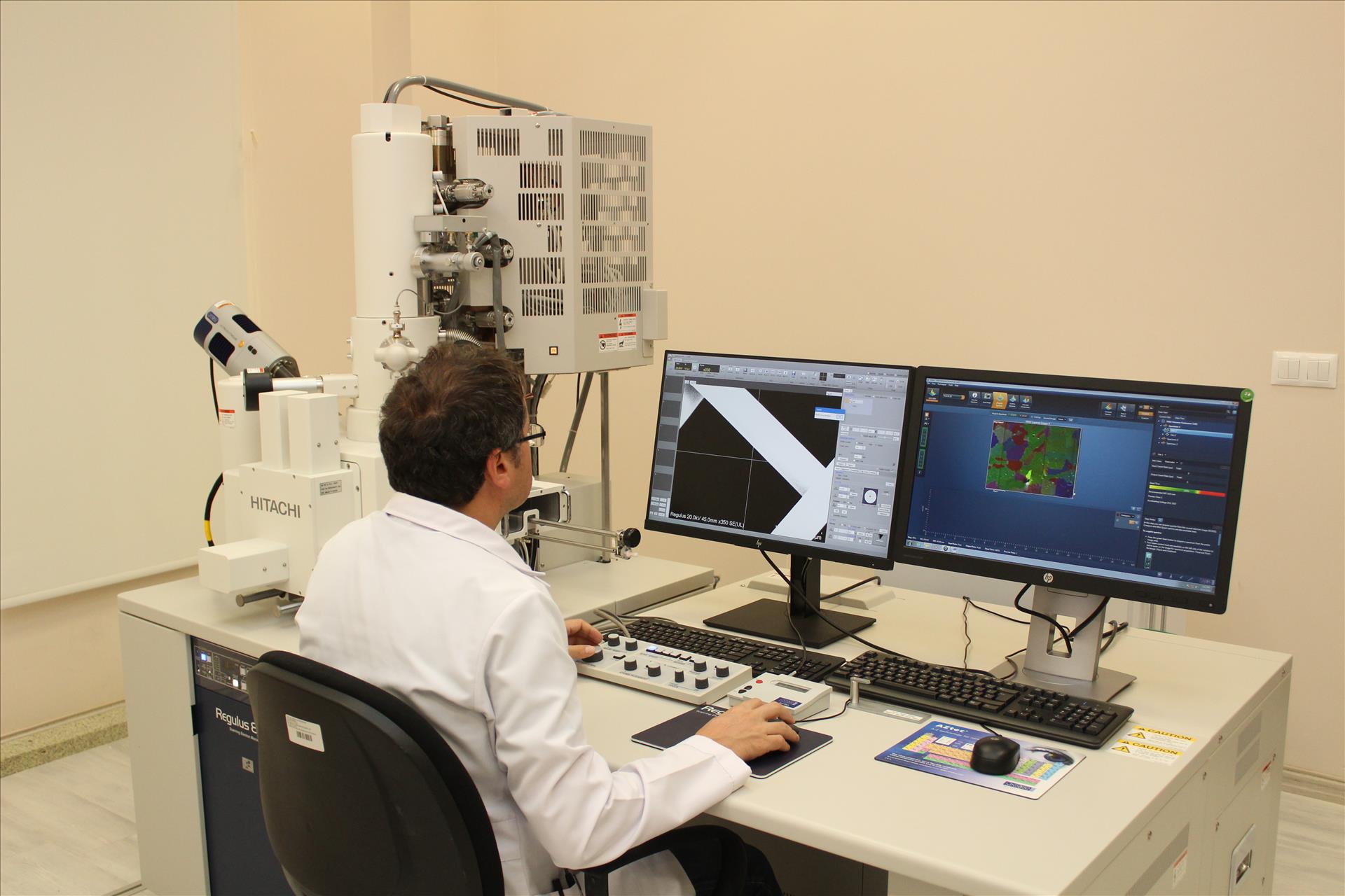
SCANNING ELECTRON MICROSCOPE (FE-SEM)
In Scanning Electron Microscopy, the electrons supplied from an electron source are directed onto the sample by means of collecting lenses in a column under vacuum.
Secondary electrons scattered from surface atoms provide information about the sample surface, while back scattered electrons are used to detect elementary differences in the sample surface by the contrast difference between regions with different chemical compositions. The back scattered electrons also provide electron back scattering diffraction (EBSD) images, for example, to help while determining the crystallographic structure. The x-rays resulting from the interaction of the electron beam with the sample are collected by an EDS detector and are used in Energy-Distributed X-Ray Spectroscopy.
Hitachi Regulus 8230 brand / model scanning electron microscope, which operates in our center, has a Cold Field Emission Electron Gun and has a resolution of 0.9 nm at 1 kV. The microscope runs in the range of 0.5 to 30 kV and the high voltage can be reduced to even 10 V. Moreover, the beam deceleration feature, thus, depending on the sample properties, provides high resolution at low voltages.
Through the EDS detector integrated into the device, the abundance of the elements in the sample can be detected and mapped, the crystallographic structure of the sample is determined by the EBSD detector, and it is possible to obtain information about the internal structures of biological samples, thin films or polymeric materials by means of the STEM detector.
In our center, Cold Field Emission SEM has a wide operating voltage range, high resolution capability at low voltages, and depending on the sample properties, has the ability to perform magnificationup to 2000k. Its lens structure and cold field emission gun provides the user high resolution imaging and high sensitivity characterization. It is equipped to meet the demands of the users in many fields such as physics, chemistry biology, medical sciences.
Device: Hitachi Regulus 8230 FE-SEM
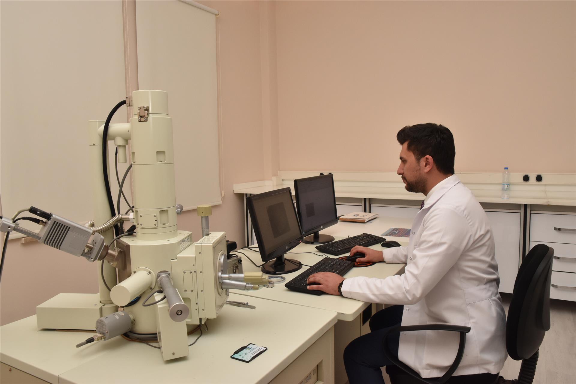
SCANNING ELECTRON MICROSCOPE (SEM)
The scanning electron microscope is a type of electron microscope that obtains images by scanning the sample surface with a focused electron beam. Electrons interact with the atoms in the sample to produce different signals containing information about the topography and composition on the sample surface. These signals are collected by the respective detectors and transferred to the computer screen and the image is obtained.
In the SEM section of the ARUM, there is a JEOL JSM 5600 LV SEM device. EDS system provides point, line and area scanning and X-ray mapping over a selected area. For insulating samples, gold palladium and carbon coating devices, and for delicate samples, a critical point dryer device are utilised in the sample preparation section. In this way, physiological, pathological or morphological changes in tissues are analysed via SEM imaging.
SEM Device: JEOL JSM 5600
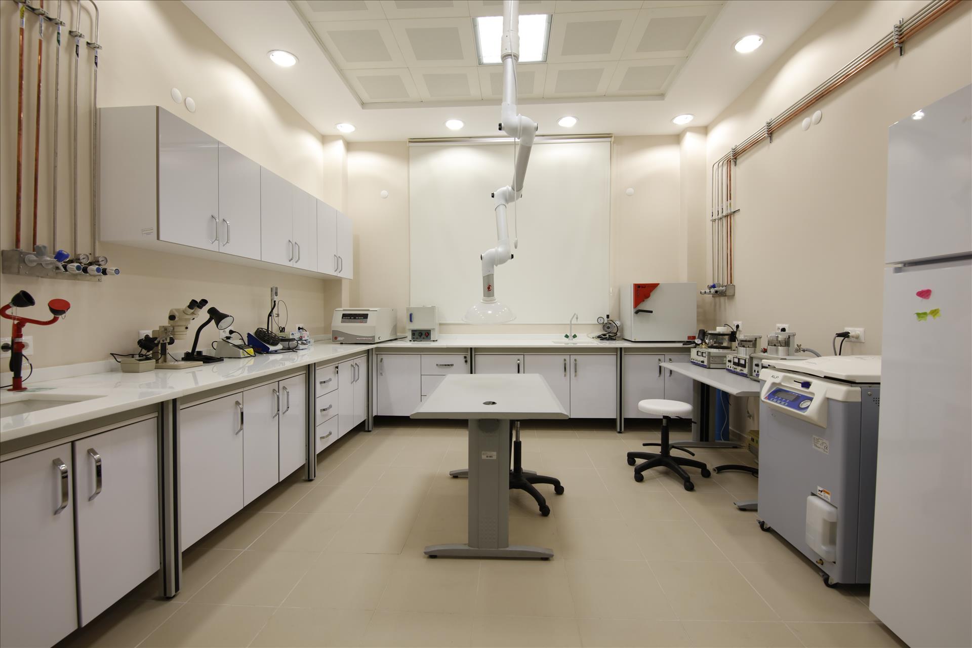
SEM SAMPLE PREPARATION
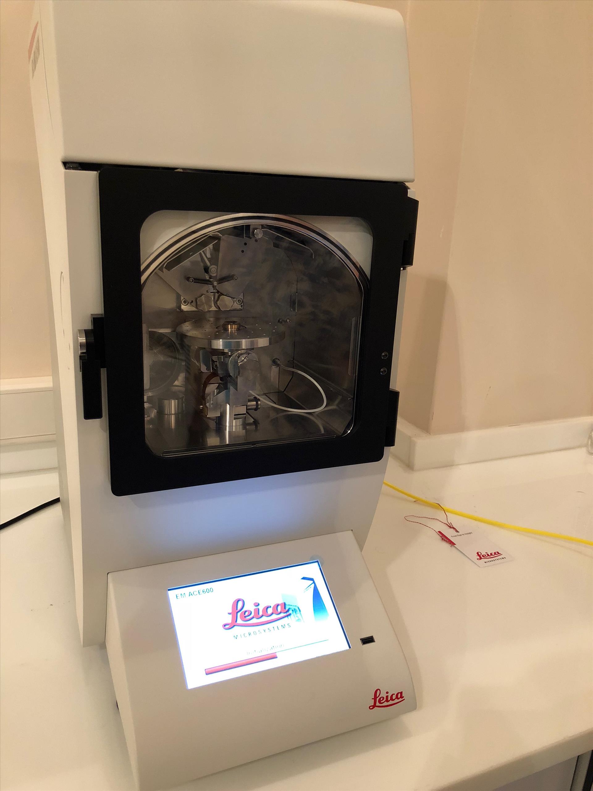
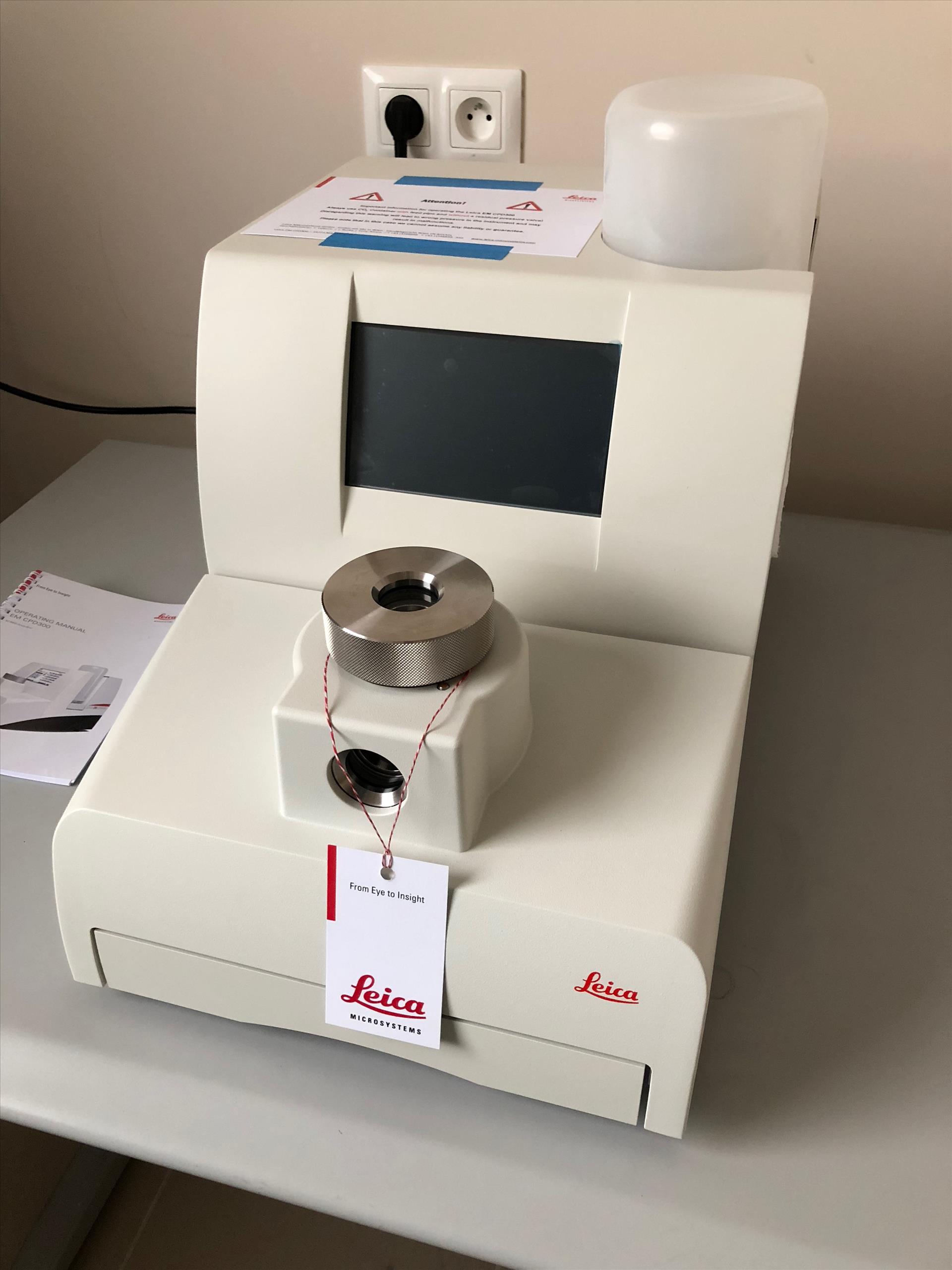
In our SEM sample preparation laboratories within the Electron Microscopy Service Unit, Leica EM CPD300 Critical Spot Drying Device is used for drying of polymeric and various materials such as airgel and hydrogel as well as biological samples. In addition, the desired non-conductive samples can be coated with gold / palladium (Au / Pd) or carbon (C) in the desired coating thickness with our Leica EM ACE600 coating device.
In addition, our SEM sample preparation laboratory provides the researcher with the necessary technical and chemical infrastructure and the necessary pre-chemical preparation and fixation processes for the biological materials required for SEM imaging.
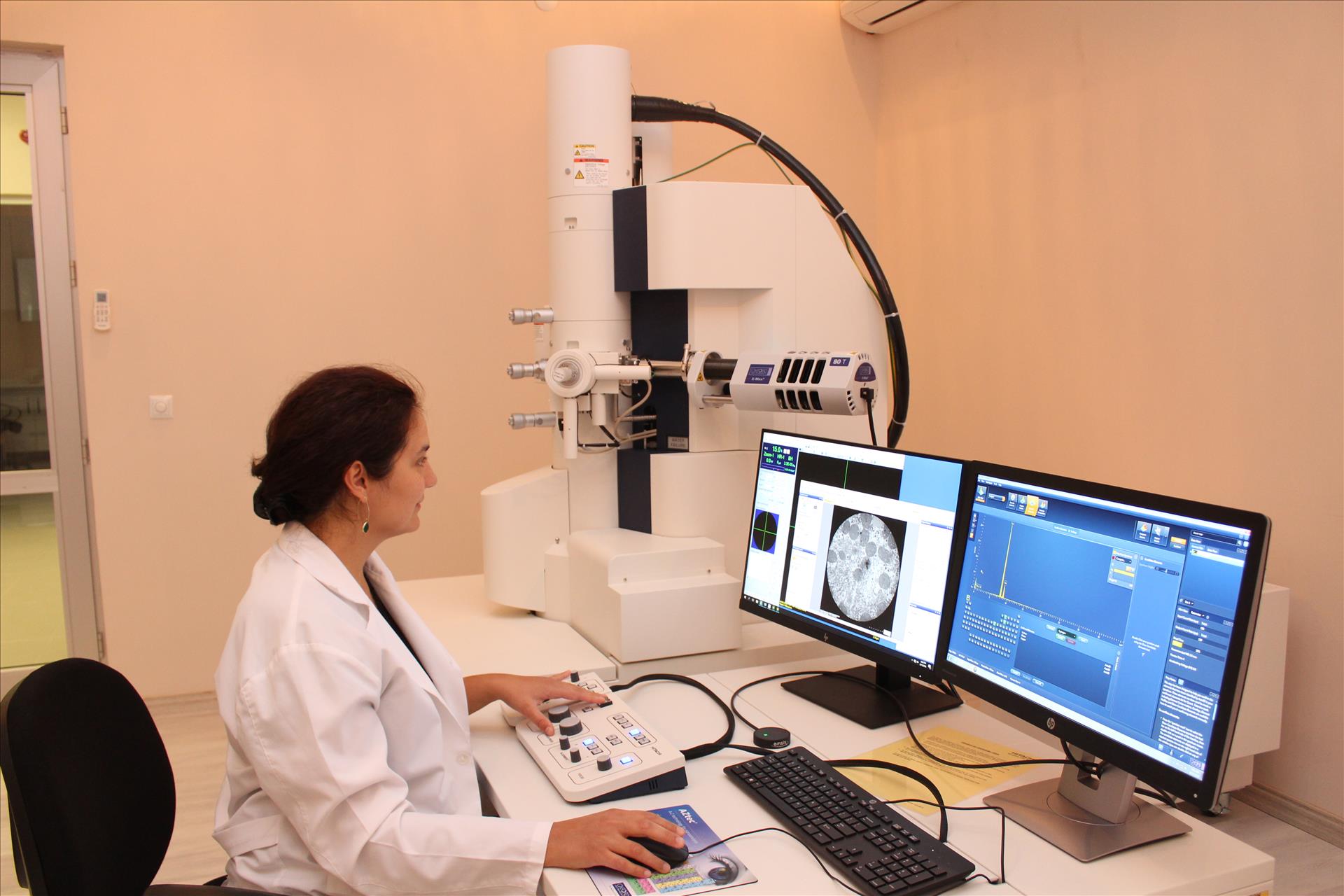
CRYO-TRANSMISSION ELECTRON MICROSCOPE (Cryo-TEM)
Hitachi HT7800 TEM brand transmission electron microscope, which is in service at our center, provides high contrast and high resolution with its multiple lens configurations and allows for many specialized applications. The device can operate up to 120 kV accelerating voltage and provides the elemental analysis of the samples with the EDS detector that can scan the Boron-Uranium (5B-92 U) elemental range. Dual-Mode lens lenses in the device provide high contrast and resolution in all areas, even in low magnifications. Furthermore, thanks to the advanced stage navigation function, the entire grid is quickly scanned and the desired zone is easily detected at the desired magnifications.
The attachments that allows the cryogenic device, thanks to our work is the first and only in Turkey in this field. The most important advantage of cryo-TEM, which is popular in the world and especially in the field of structural biology, is that it allows the biomolecules to be seen in a lot of processes which cannot be observed before by freezing while they are in motion. On the other hand, in conventional TEM examinations, the problems such as radiation damage and high vacuum in the column, for example, are greatly exceeded by cryo-TEMs. Thus, it is possible to examine the samples with much less damage.
Analyzes:
- Ultrastructural investigations of all biological samples,
- Chemical and structural characterization of nanomaterials,
- Structure investigations of semiconductors,
- Morphological researches,
- Polymeric and glassy materials,
- Energy technology studies,
- Lightening of 3D structures of cells and tissues,
- Investigation of viruses and macromolecular complexes.
CRYO-TEM Device: Hitachi HT7800
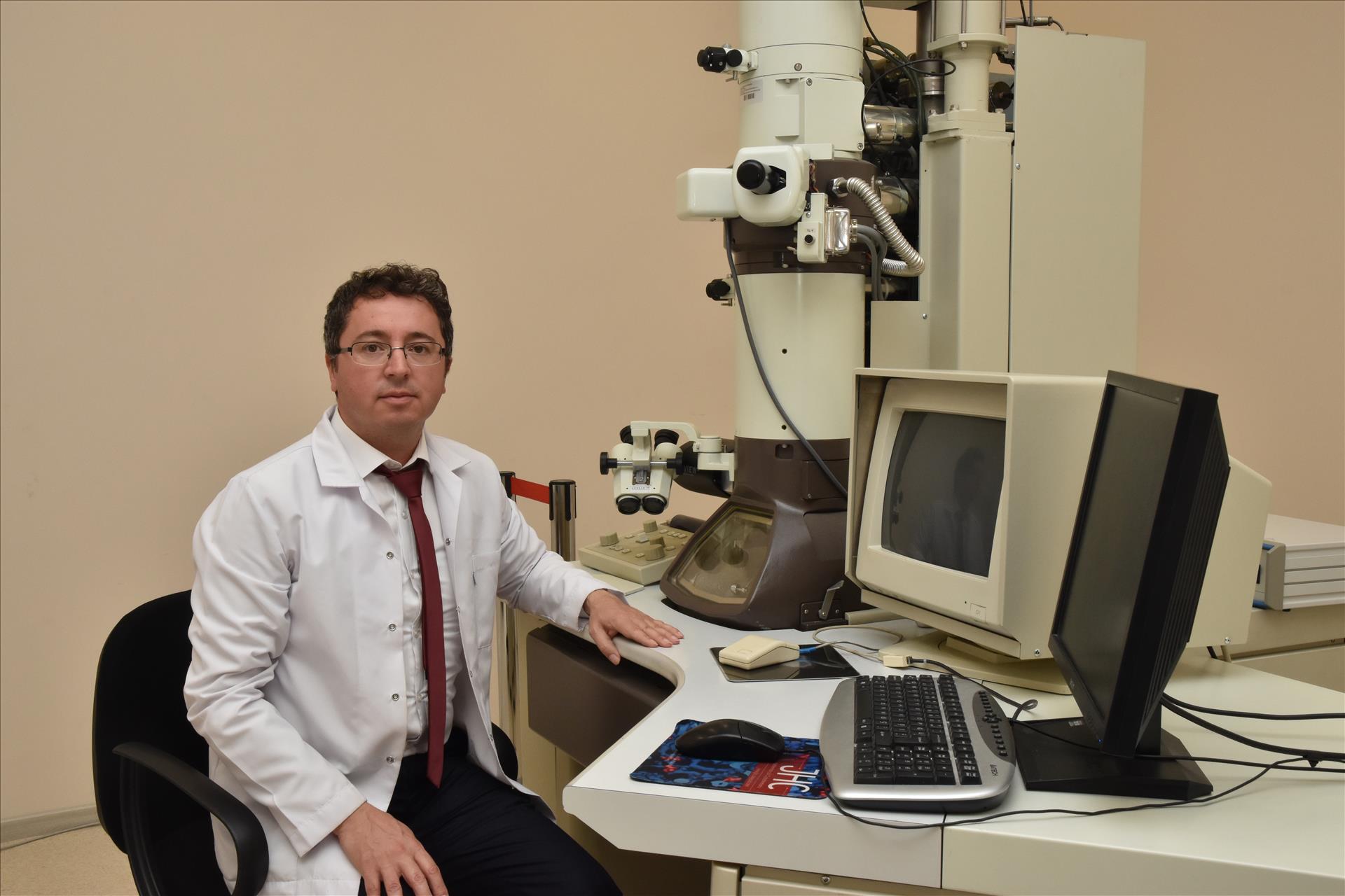
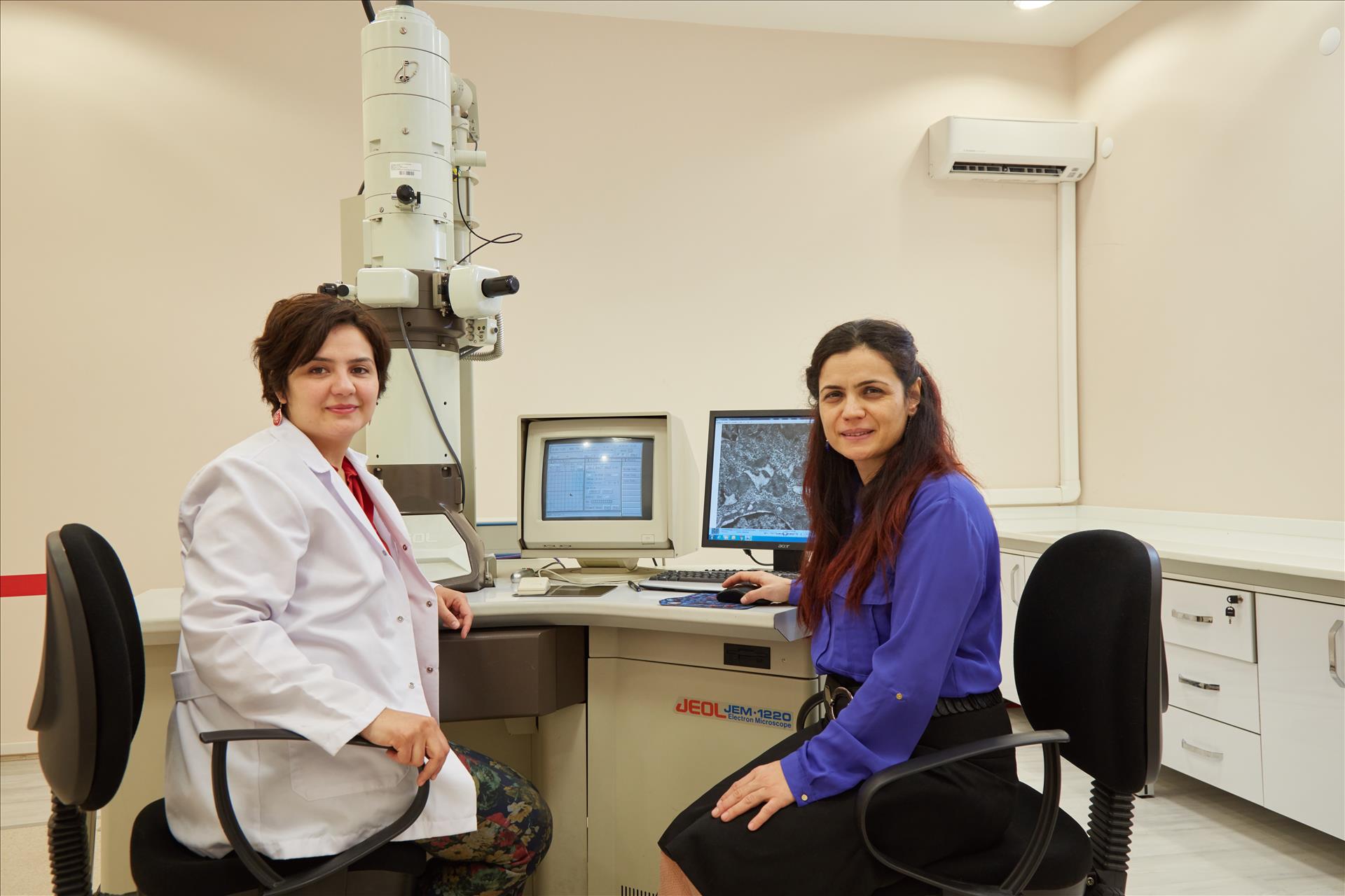
TRANSMISSION ELECTRON MICROSCOPE (TEM)
The Transmission Electron Microscope (TEM) is based on the principle of displaying high-energy electrons passed through a very fine sample. The image resulting from the interaction of the electrons with the sample is enlarged and the fluorescence screen focuses on a sensor, such as a photographic film layer or a CCD camera.
ARUM Central Laboratory TEM section contains JEOL 1220 JEM TEM device. In our department, routine electron microscopic follow-up procedures including tissue follow-up, sectioning and staining procedures are also carried out for the preparation of biological samples.
Thanks to this technique, morphological, structural and elemental information can be obtained from low-magnification to high-magnification (50x-200.000x). In the fields of biology, medicine and basic science, the analysis of the surface and fine structures of the sample in high resolution and contrast, morphological research, interpretation of structure-function relations can be done.
TEM Device: JEOL JEM 1220
Analysis with TEM;
- Detailed view of the surface and fine structures of the sample
- Morphological researches,
- Interpretation of structure-function relations
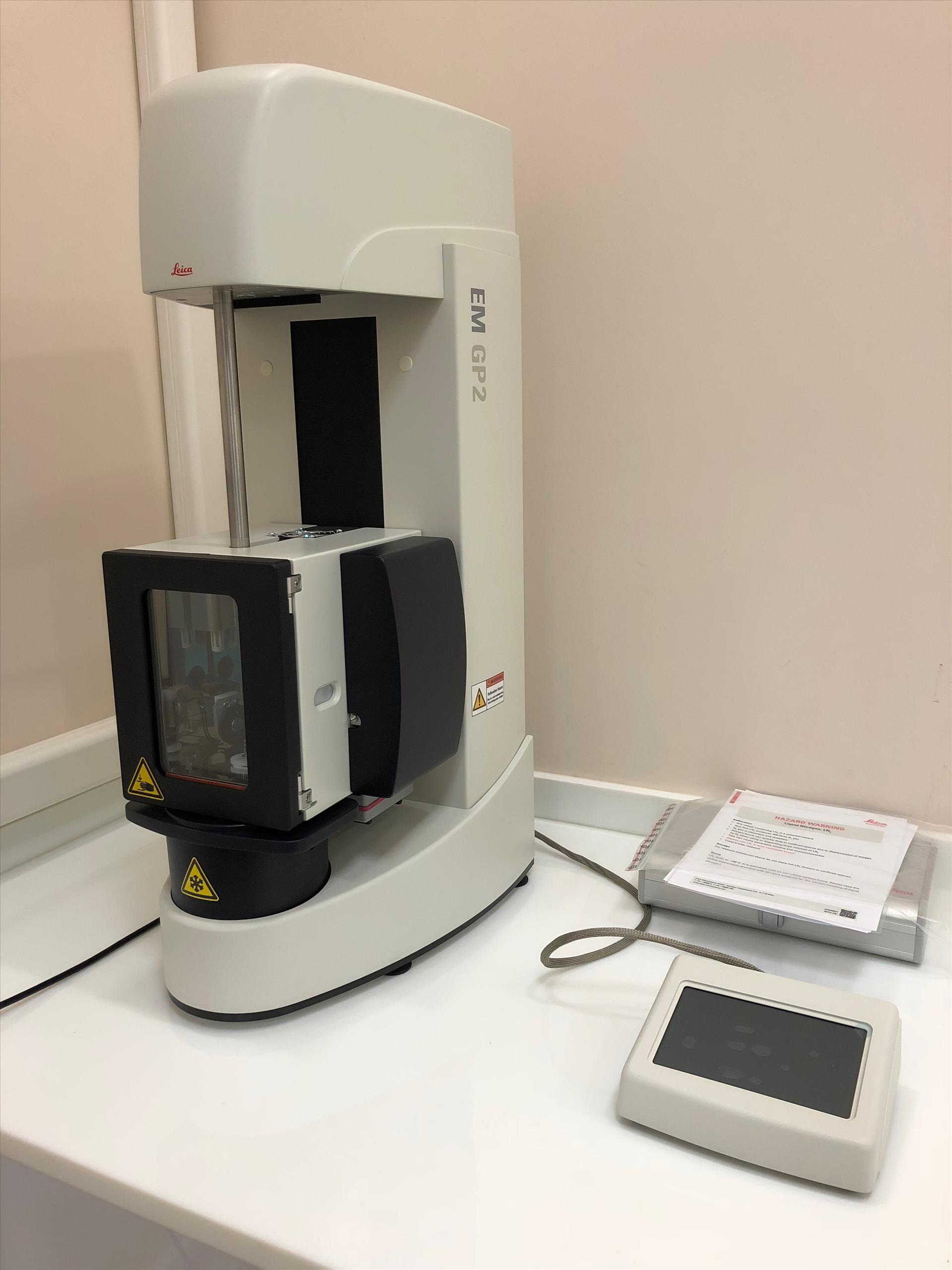
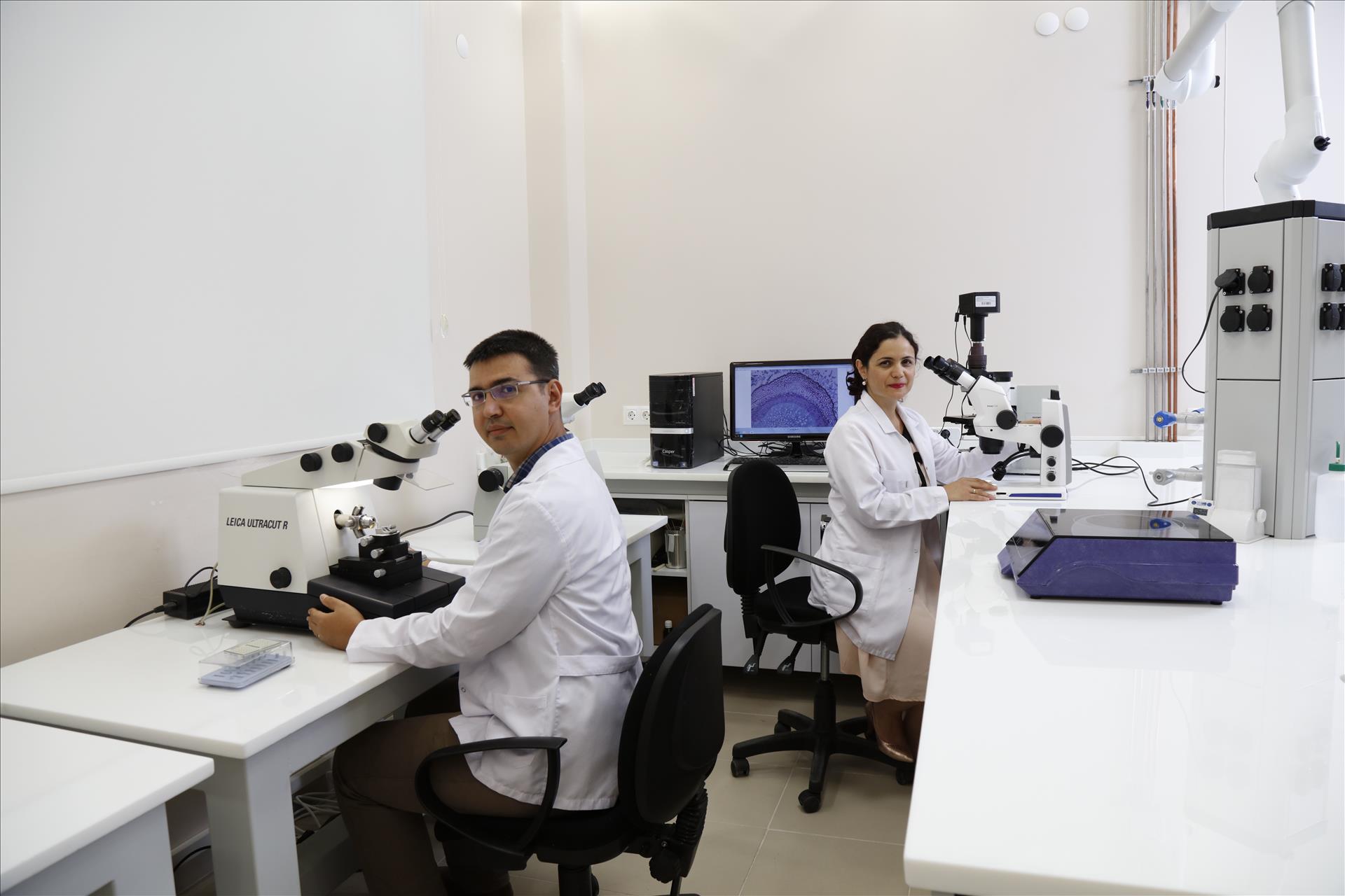
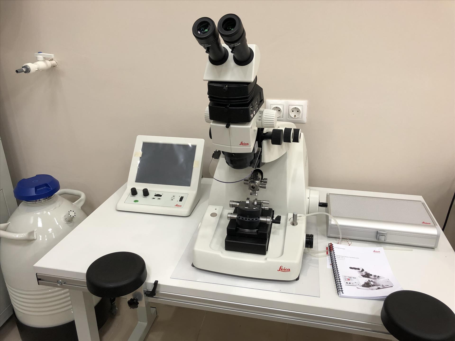
KRIO-TEM SAMPLE PREPARATION
The TEM Sample Preparation Laboratory within the Electron Microscopy Unit has successfully carried out the chemical preparation and fixation processes prior to the TEM imaging of biological materials with its expert staff and technical equipment. Our laboratory infrastructure has been extended with appropriate equipment and put into service for our researchers in cryogenic TEM studies.
The Leica EM UC7 brand / model ultramicrotome in the TEM preparation laboratory is an ergonomic design that provides easy and easy removal of semi-thin and full thin sections of various biological and industrial samples. The device is equipped with attachments that can be used in both room conditions and cryo temperatures. User, sample, blade and storage parameters can be easily loaded with a USB. The device has three independent LED lamps with built-in brightness control and additional LED spotlights to enhance vision. A cryo-chember can be mounted to the device within a few minutes to convert the ultramicrotome into a cryo-ultramicrotome and cryo-sections (-15 ° to -185 ° C) can be removed. Biological and industrial sample sections obtained by ultramicrotome in both options can be used for examination in microscopes such as TEM, SEM, AFM and LM.
The Leica EM GP2 device in our TEM preparation laboratory is used to prepare vitrified samples for cryo-TEM analysis. Industrial samples in biological or aqueous / inorganic solvents with the aid of the device can be prepared by immersion-freezing technique. In this method, the sample in the liquid is pipetted onto the EM gradient and the excess liquid is removed. The grid immersed in a cryogen, such as liquid ethane, is then directly cryopreserved and examined under cryo-conditions. Prior to freezing the sample is protected in a chamber, temperature (+ 4 ° C and + 60 ° C) and humidity controlled (99%).
Viruses, proteins, macromolecules complexes and industrial emulsions can be prepared by this technique. This ensures that the biomolecules frozen in motion are visualized with minimal damage and maximum resolution.