SCANNING ELECTRON MICROSCOPE (SEM)
The scanning electron microscope is a type of electron microscope that obtains images by scanning the sample surface with a focused electron beam. Electrons interact with the atoms in the sample to produce different signals containing information about the topography and composition on the sample surface. These signals are collected by the respective detectors and transferred to the computer screen and the image is obtained.
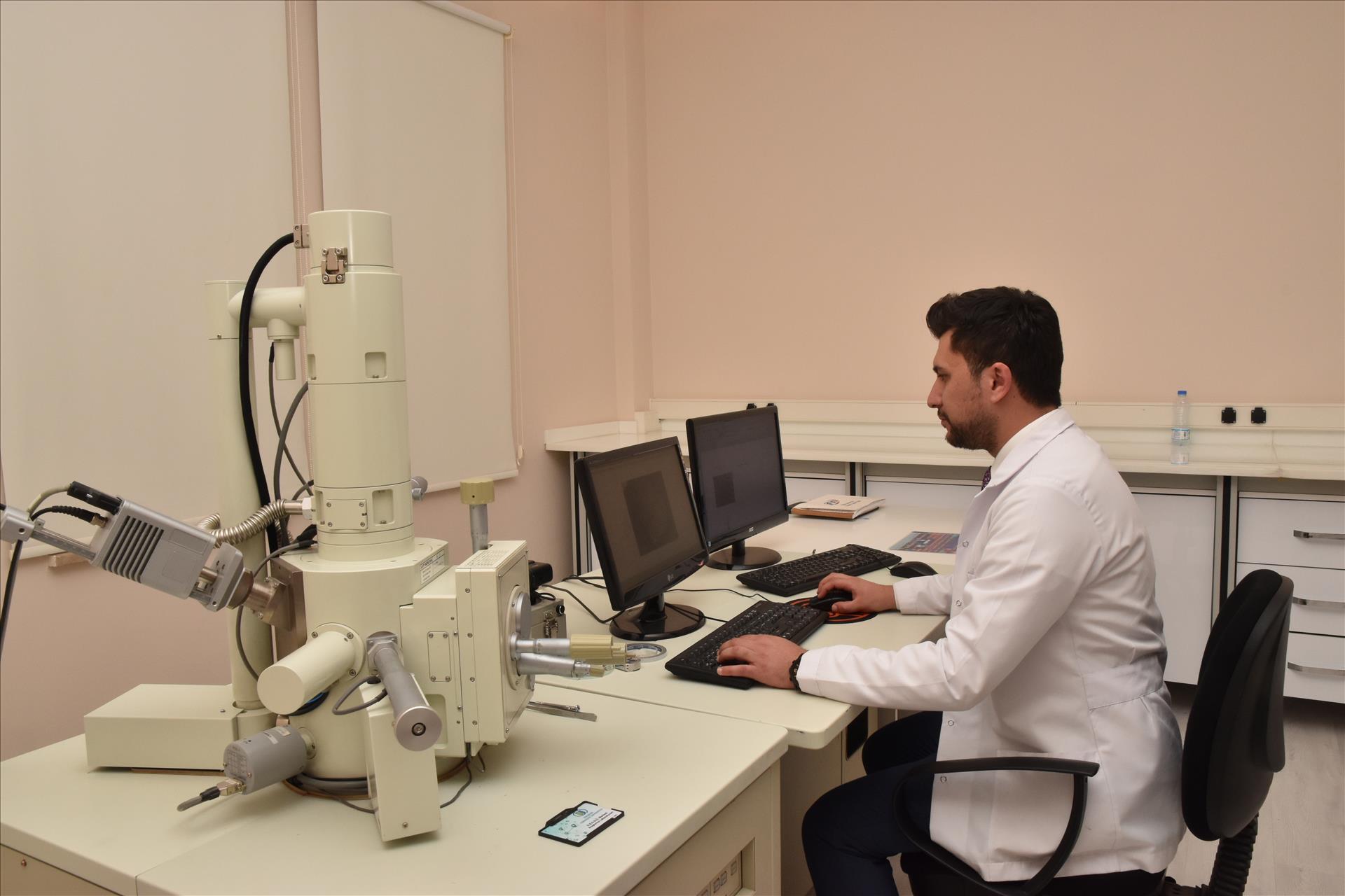
In the SEM section of the ARUM Center laboratory there is the SEM device of the JEOL JSM 5600. A designated point, line and area scanning and selected area X-ray mapping are carried out with EDS system. For the preparation of samples for the analysis of insulating samples, there is a gold palladium coating device which runs under high vacuum. There is also a point dryer for biological samples. In this way, physiological or pathological tissues in tissues, or morphological changes that occur with experimental methods are analyzed.
SEM Device: JEOL JSM 5600