TRANSMISSION ELECTRON MICROSCOPE (TEM)
The Transmission Electron Microscope (TEM) is based on the principle of displaying high-energy electrons passed through a very fine sample. The image resulting from the interaction of the electrons with the sample is enlarged and the fluorescence screen focuses on a sensor, such as a photographic film layer or a CCD camera.
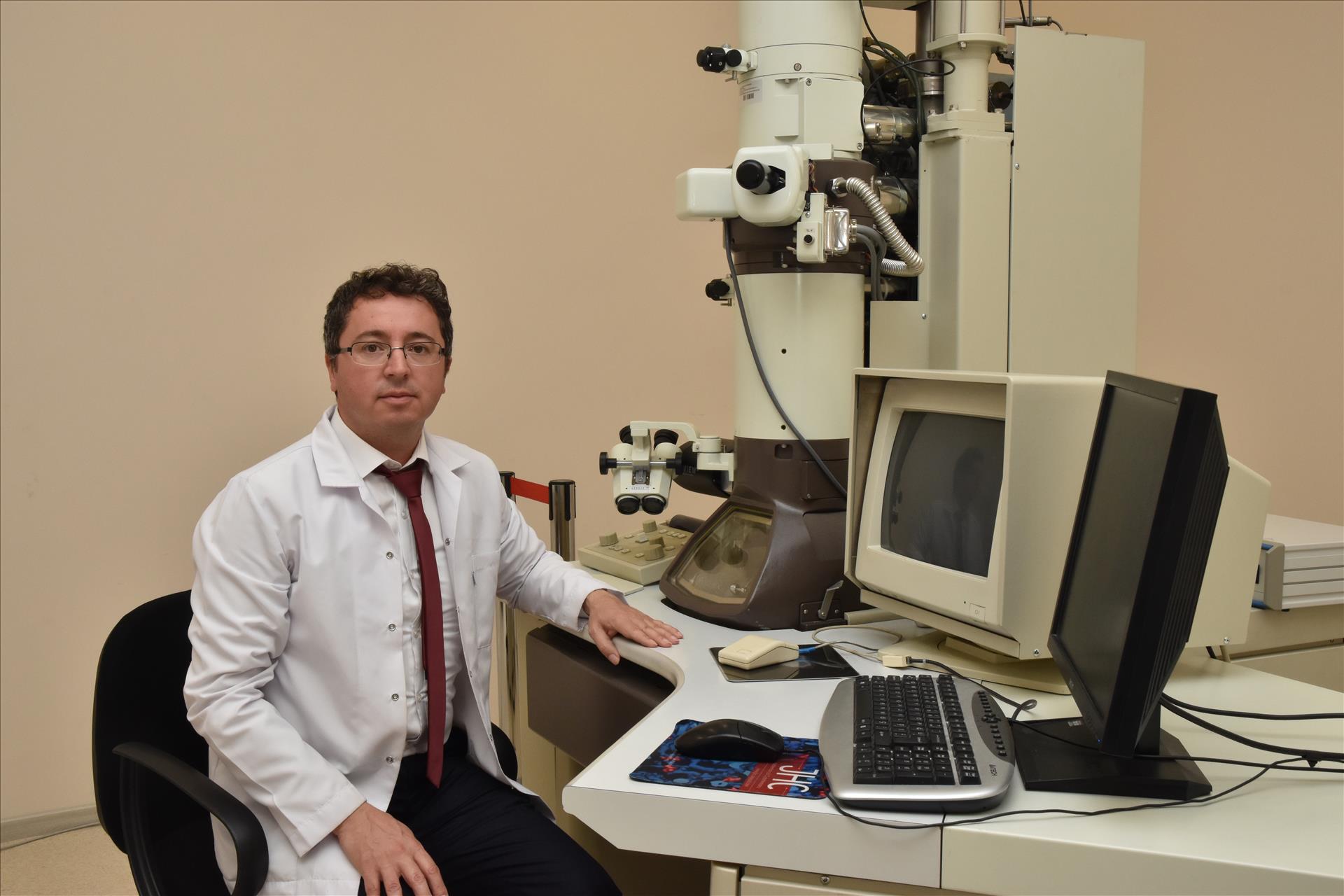
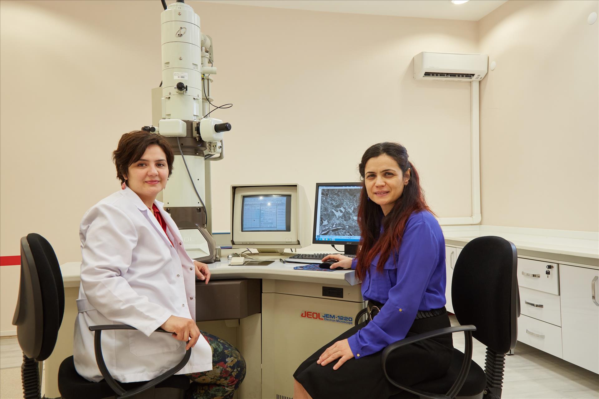
ARUM Central Laboratory TEM section contains JEOL 1220 JEM TEM device. In our department, routine electron microscopic follow-up procedures including tissue follow-up, sectioning and staining procedures are also carried out for the preparation of biological samples.
Thanks to this technique, morphological, structural and elemental information can be obtained from low-magnification to high-magnification (50x-200.000x). In the fields of biology, medicine and basic science, the analysis of the surface and fine structures of the sample in high resolution and contrast, morphological research, interpretation of structure-function relations can be done.
TEM Device: JEOL JEM 1220
Analysis with TEM;
- Detailed view of the surface and fine structures of the sample
- Morphological researches,
- Interpretation of structure-function relations