CRYO-TEM SAMPLE PREPARATION
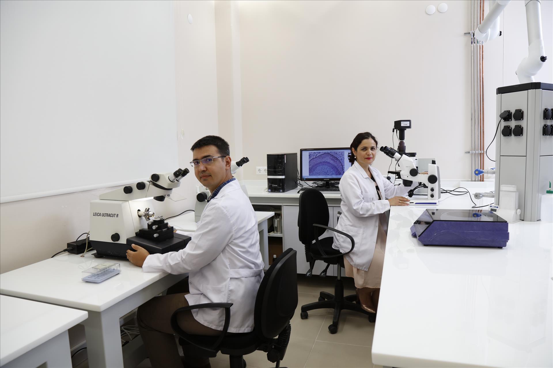
The TEM Sample Preparation Laboratory within the Electron Microscopy Unit has successfully carried out the chemical preparation and fixation processes prior to the TEM imaging of biological materials with its expert staff and technical equipment. Our laboratory infrastructure has been extended with appropriate equipment and put into service for our researchers in cryogenic TEM studies.
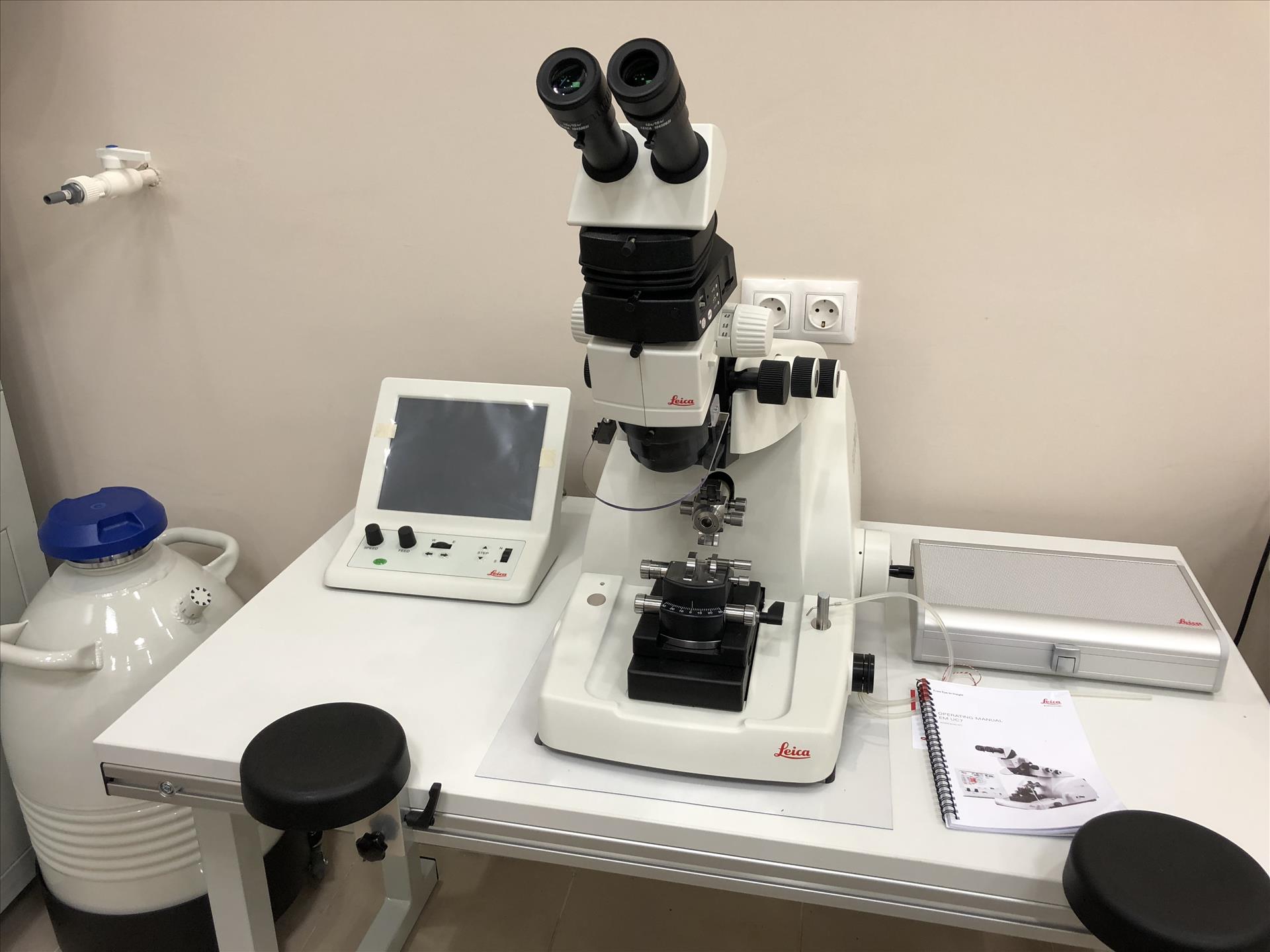
The Leica EM UC7 brand / model ultramicrotome in the TEM preparation laboratory is an ergonomic design that provides easy and easy removal of semi-thin and full thin sections of various biological and industrial samples. The device is equipped with attachments that can be used in both room conditions and cryo temperatures. User, sample, blade and storage parameters can be easily loaded with a USB. The device has three independent LED lamps with built-in brightness control and additional LED spotlights to enhance vision. A cryo-chember can be mounted to the device within a few minutes to convert the ultramicrotome into a cryo-ultramicrotome and cryo-sections (-15 ° to -185 ° C) can be removed. Biological and industrial sample sections obtained by ultramicrotome in both options can be used for examination in microscopes such as TEM, SEM, AFM and LM.
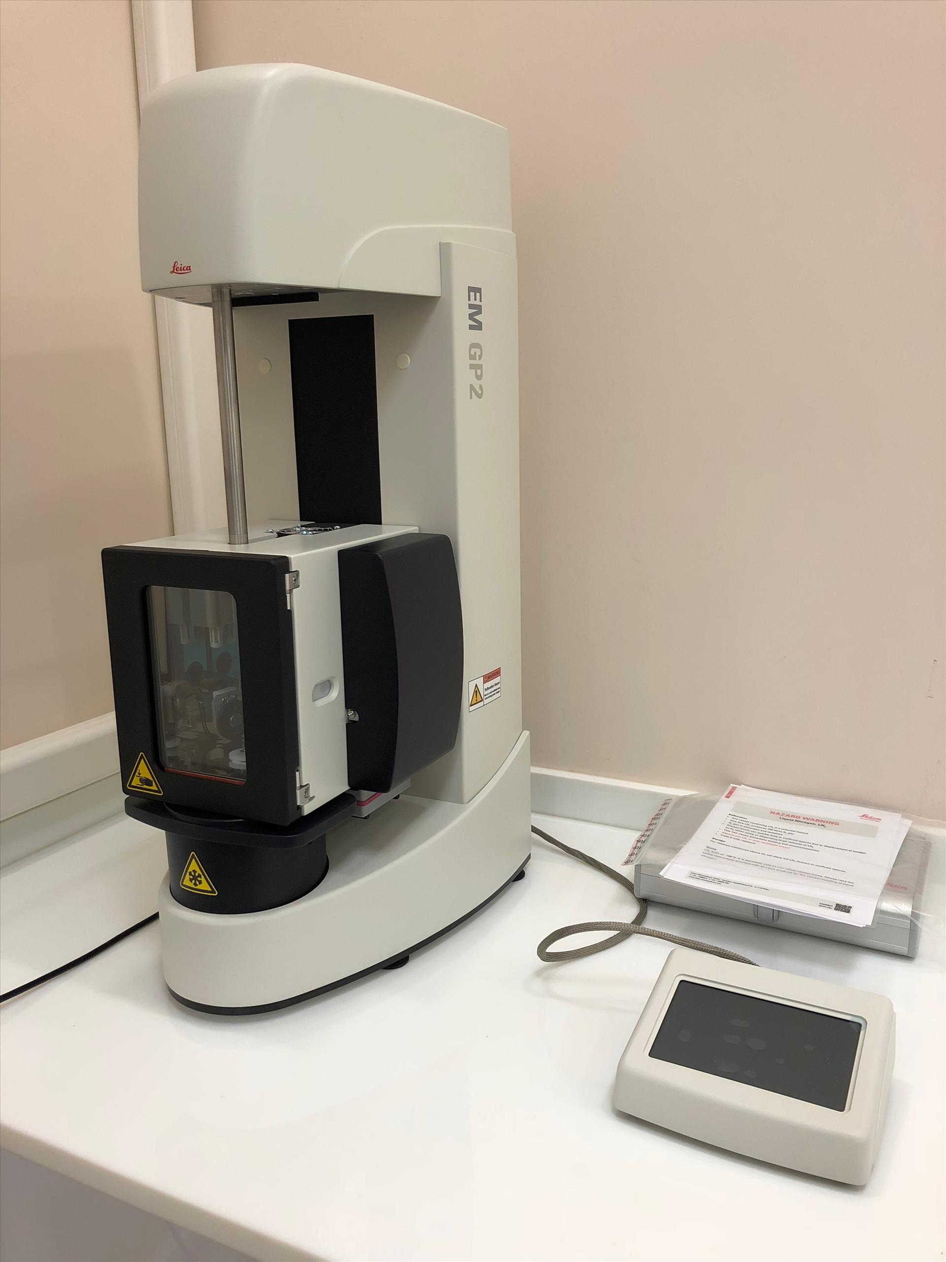
The Leica EM GP2 device in our TEM preparation laboratory is used to prepare vitrified samples for cryo-TEM analysis. Industrial samples in biological or aqueous / inorganic solvents with the aid of the device can be prepared by immersion-freezing technique. In this method, the sample in the liquid is pipetted onto the EM gradient and the excess liquid is removed. The grid immersed in a cryogen, such as liquid ethane, is then directly cryopreserved and examined under cryo-conditions. Prior to freezing the sample is protected in a chamber, temperature (+ 4 ° C and + 60 ° C) and humidity controlled (99%).
Viruses, proteins, macromolecules complexes and industrial emulsions can be prepared by this technique. This ensures that the biomolecules frozen in motion are visualized with minimal damage and maximum resolution.