CONFOCAL LASER SCANNING MICROSCOPE (CLSM)
A confocal microscope is a imaging technique using a small planar orifice called pinhole to eliminate the unfocused light in the samples that are thicker than the point lighting and focal plane to increase the optical resolution and visibility of a microscopic image. It provides clearance at different levels within the section thickness and provides optical sections finer than the sections. The most important advantage is that it can collect light from a single plan. In this technique, three-dimensional structures of images obtained by studying thick tissue sections, developing embryos, or whole cell samples marked with fluorescent or reflective probes can be determined. It can be applied to single or multiple marked samples. The confocal microscope also gains importance in the study of semiconductors and material science.
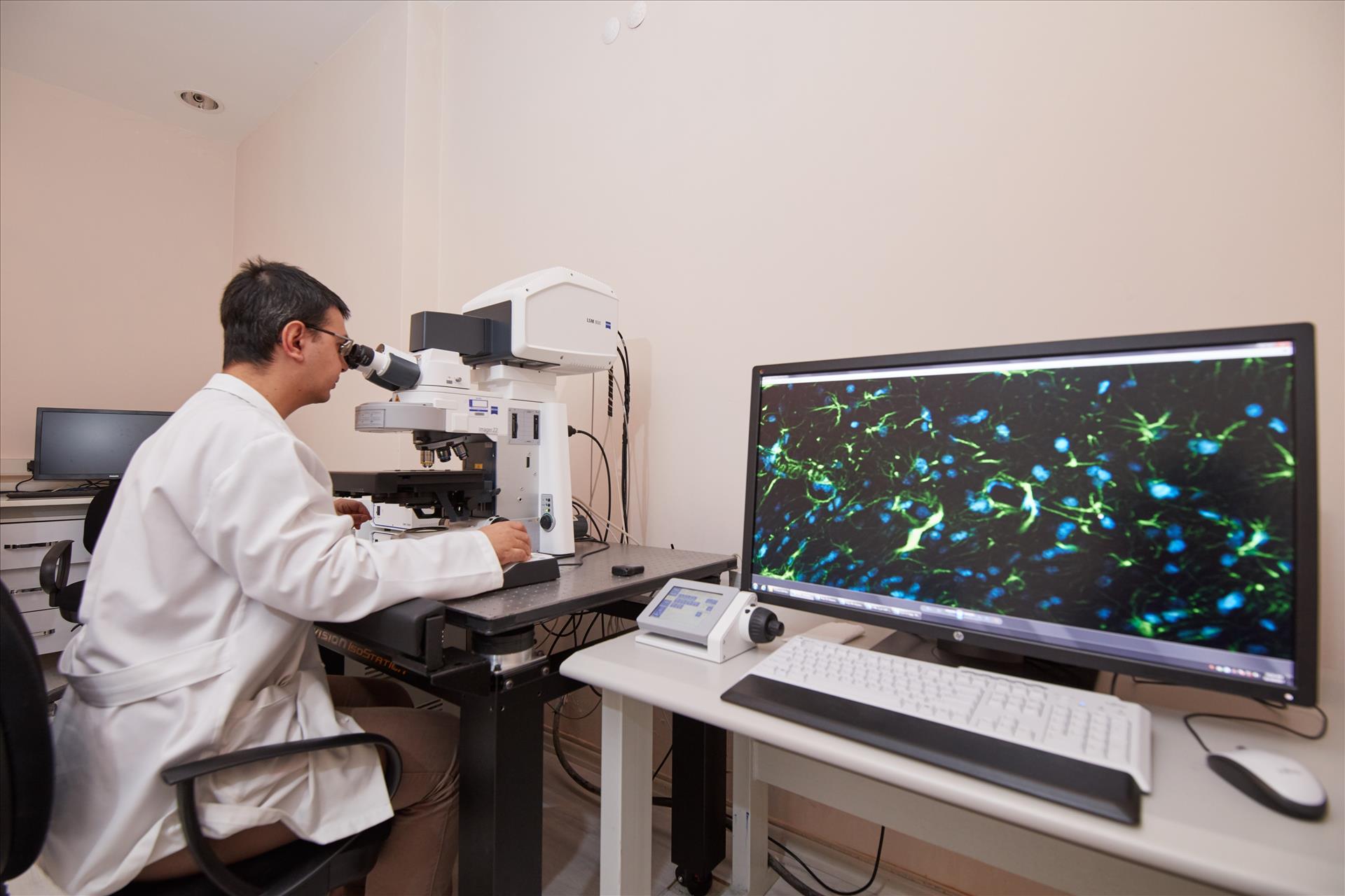
ZEISS LSM800 brand laser scanning confocal microscope and images analysis, processing and three-dimensional configuration is required for the software package is available. The microscope system includes diode laser sources (405 nm, 488 nm, 561 nm and 640 nm).
CLSM Device: ZEISS LSM 800
Analysis with CLSM;
- High-resolution and high-visibility 3D imaging,
- Fixed cell or life instantly viewing,
- Morphological imaging,
- Examination of developing embryos
- Control of biofilms
- 2B-5B (xyztλ) display,
- Cell culture and nerve cell imaging,
- High resolution and multicolor fluorescence analysis,
- Complex accelerated experiment shots.