RAMAN SPECTROMETER
Raman spectroscopy works according to the method by which the bonds in the molecule inflate the light that is lowered on these bonds inexorably. Raman spectrometry and microscopes are a sensitive measurement method that can be used in all studies in a wide range of sciences such as basic sciences, pharmacy, medicine and many other engineering disciplines.
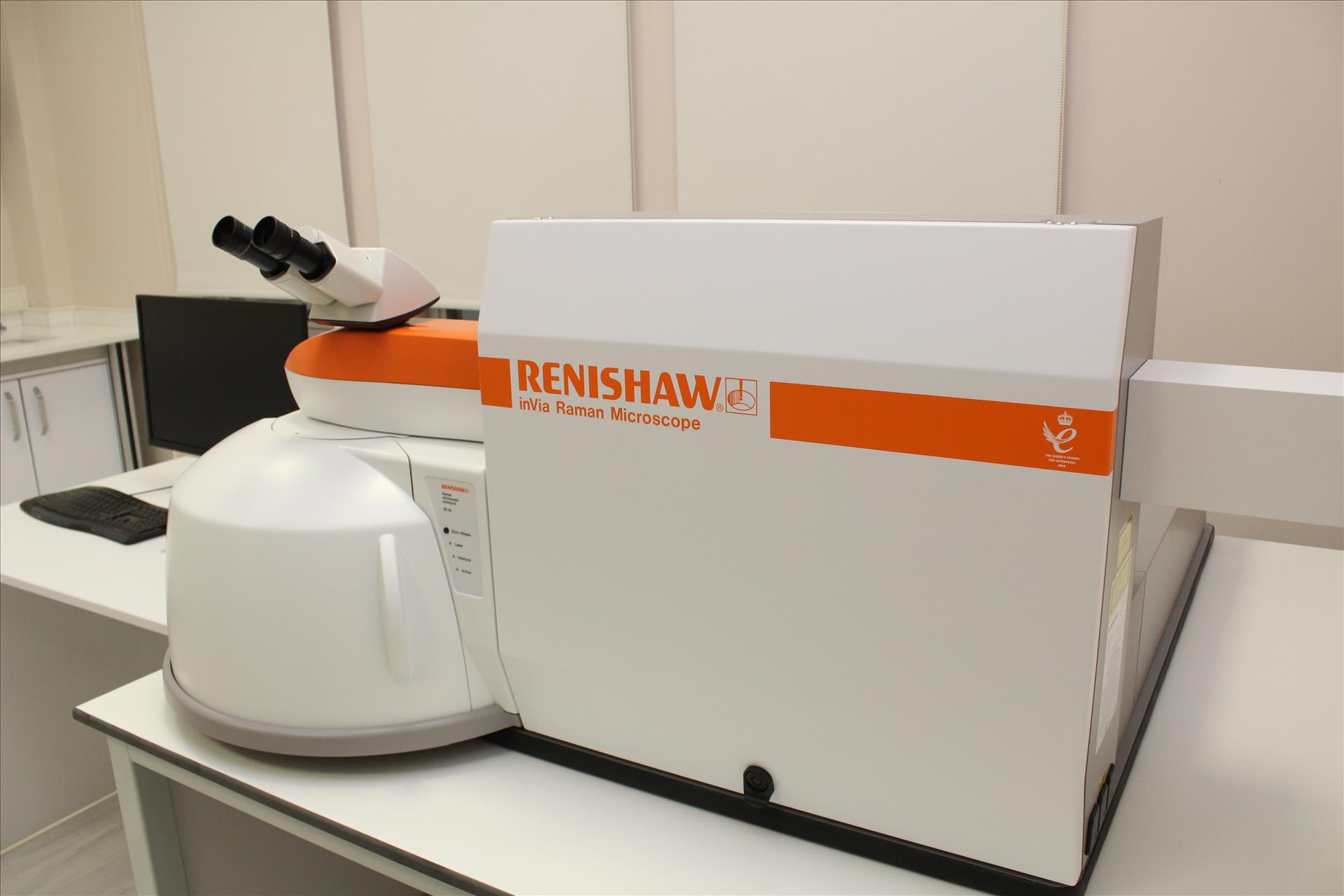
In the spectroscopy section of ARUM laboratory there are renishaw brand Raman spectroscopy. It is important in technological and scientific research considering its fast and sensitive, low sample requirements, ease of use and non-destructive properties.
By microscopic imaging, both the sample surface can be visualized and the point and area analysis can be performed from the desired surface area. In addition, field mapping and profiling can be done in 3 dimensions with our Raman system.
Raman Device: Renishaw inVia Raman Microscope
Lasers: 532 nm, 735 nm
Analysis with Raman spectroscopy;
- Determination of chemical and structural information of samples,
- In the analysis of polymer mixtures,
- In stress analysis of fibers,
- In determination of crystallinity levels,
- Micrometer-based spatial resolution of drug formulations,
- In the examination of thermal behavior of drugs with advanced temperature control,
- In determining the homogeneity of the mixture,
- Analysis of API and metabolites in cells and tissues,
- In determining the number, defects, additives and voltages of carbon based materials,
- In determining the diameter and function of carbon nanotube (CNT),
- In stress / stress analysis,
- In determination of additive concentrations,
- Thin film thickness determination,
- In determining the crystal structure type and orientation,
- In determination of crystal quality,
- In the determination of homogeneity and purity,
- To distinguish cancerous cells from normal cells,
- To distinguish stem cells from differentiated cells,
- In defining different sub states in a cell population,
- To reliably mark the boundaries of the anatomical layers in the tissues and to define them objectively,
- It can be used in the identification of chemical changes that occur in tissues but do not reveal themselves as morphological changes.